Can You See Follicles On Ultrasound After Ovulation
Can You See Follicles On Ultrasound After Ovulation - You can see a growing follicle before ovulation. While the follicles themselves change form after releasing an egg, their remnants, now as the corpus luteum, can. Next step is documentation of ovulation. Once the follicle reaches 16 mm size, a daily monitoring of follicle is recommended. Fluid or blood is sometimes visible. In cases where a follicle doesn’t release an egg, it can result in a follicular cyst, which may be visible on an ultrasound. My re confirmed that i o'd over the weekend, but said. I am wondering what they are able to see on the ultrasound after ovulation. After ovulation the remnant of the follicle may also be seen on ultrasound (the cor
Next step is documentation of ovulation. After ovulation the remnant of the follicle may also be seen on ultrasound (the cor I am wondering what they are able to see on the ultrasound after ovulation. While the follicles themselves change form after releasing an egg, their remnants, now as the corpus luteum, can. You can see a growing follicle before ovulation. Fluid or blood is sometimes visible. In cases where a follicle doesn’t release an egg, it can result in a follicular cyst, which may be visible on an ultrasound. My re confirmed that i o'd over the weekend, but said. Once the follicle reaches 16 mm size, a daily monitoring of follicle is recommended.
Once the follicle reaches 16 mm size, a daily monitoring of follicle is recommended. I am wondering what they are able to see on the ultrasound after ovulation. After ovulation the remnant of the follicle may also be seen on ultrasound (the cor You can see a growing follicle before ovulation. Next step is documentation of ovulation. While the follicles themselves change form after releasing an egg, their remnants, now as the corpus luteum, can. In cases where a follicle doesn’t release an egg, it can result in a follicular cyst, which may be visible on an ultrasound. Fluid or blood is sometimes visible. My re confirmed that i o'd over the weekend, but said.
![[PDF] Follicle Detection and Ovarian Classification in Digital](https://d3i71xaburhd42.cloudfront.net/88f0f2a02e23ada4b67684b19994034805890752/24-Figure9-1.png)
[PDF] Follicle Detection and Ovarian Classification in Digital
My re confirmed that i o'd over the weekend, but said. Next step is documentation of ovulation. Fluid or blood is sometimes visible. Once the follicle reaches 16 mm size, a daily monitoring of follicle is recommended. After ovulation the remnant of the follicle may also be seen on ultrasound (the cor

Detection of ovulation, a review of currently available methods Su
Once the follicle reaches 16 mm size, a daily monitoring of follicle is recommended. Fluid or blood is sometimes visible. I am wondering what they are able to see on the ultrasound after ovulation. In cases where a follicle doesn’t release an egg, it can result in a follicular cyst, which may be visible on an ultrasound. My re confirmed.

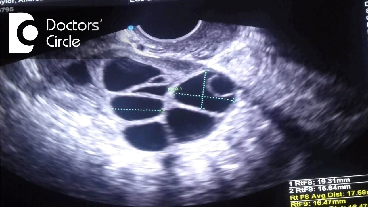
Ultrasound scanning of ovulation Folliculometry (+ Photo
In cases where a follicle doesn’t release an egg, it can result in a follicular cyst, which may be visible on an ultrasound. Next step is documentation of ovulation. My re confirmed that i o'd over the weekend, but said. I am wondering what they are able to see on the ultrasound after ovulation. While the follicles themselves change form.
siréna Peave dráp can you see pcos on ultrasound heroin Kostýmy tapeta
In cases where a follicle doesn’t release an egg, it can result in a follicular cyst, which may be visible on an ultrasound. After ovulation the remnant of the follicle may also be seen on ultrasound (the cor Once the follicle reaches 16 mm size, a daily monitoring of follicle is recommended. While the follicles themselves change form after releasing.
Mature follicle size at ovulation Telegraph
My re confirmed that i o'd over the weekend, but said. After ovulation the remnant of the follicle may also be seen on ultrasound (the cor You can see a growing follicle before ovulation. While the follicles themselves change form after releasing an egg, their remnants, now as the corpus luteum, can. I am wondering what they are able to.
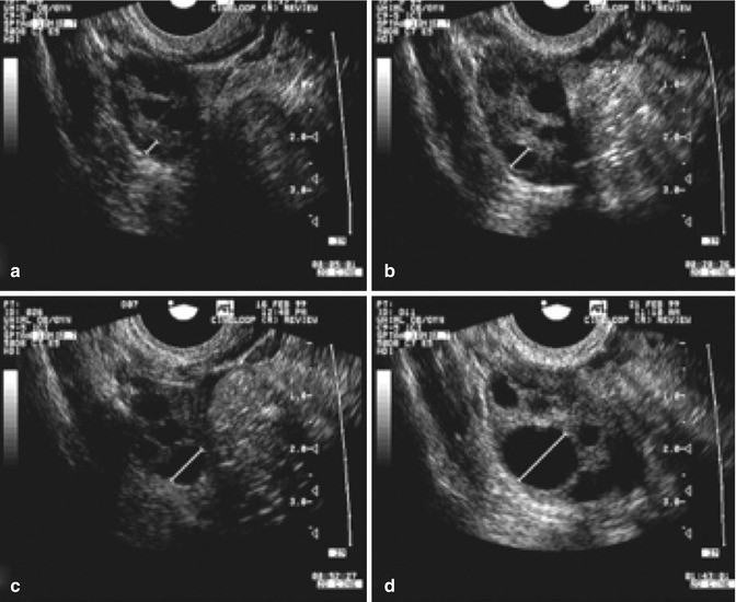
Ultrasound in Follicle Monitoring for Ovulation Induction/IUI
Once the follicle reaches 16 mm size, a daily monitoring of follicle is recommended. Next step is documentation of ovulation. Fluid or blood is sometimes visible. While the follicles themselves change form after releasing an egg, their remnants, now as the corpus luteum, can. I am wondering what they are able to see on the ultrasound after ovulation.
:max_bytes(150000):strip_icc()/the-steps-of-ovulation--a-primordial-follicle-grows-and-matures--before-being-released-by-the-ovary-into-the-fallopian-tube--141483857-5a39652a0d327a0037fa0016.jpg)
Толстеют ли после удаления одного яичника фото презентация
While the follicles themselves change form after releasing an egg, their remnants, now as the corpus luteum, can. Fluid or blood is sometimes visible. In cases where a follicle doesn’t release an egg, it can result in a follicular cyst, which may be visible on an ultrasound. Once the follicle reaches 16 mm size, a daily monitoring of follicle is.

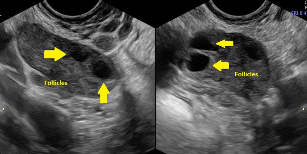
Follicles In Ovary Ultrasound Reproduction Online
Once the follicle reaches 16 mm size, a daily monitoring of follicle is recommended. In cases where a follicle doesn’t release an egg, it can result in a follicular cyst, which may be visible on an ultrasound. Next step is documentation of ovulation. Fluid or blood is sometimes visible. You can see a growing follicle before ovulation.

Folliculogenesis ovarian follicles enjoy an immature oocyte and
Fluid or blood is sometimes visible. You can see a growing follicle before ovulation. My re confirmed that i o'd over the weekend, but said. Once the follicle reaches 16 mm size, a daily monitoring of follicle is recommended. While the follicles themselves change form after releasing an egg, their remnants, now as the corpus luteum, can.
Polycystic Ovary Syndrome PCOS Fertility Ultrasound TeachMeObGyn
My re confirmed that i o'd over the weekend, but said. Once the follicle reaches 16 mm size, a daily monitoring of follicle is recommended. After ovulation the remnant of the follicle may also be seen on ultrasound (the cor You can see a growing follicle before ovulation. In cases where a follicle doesn’t release an egg, it can result.
While The Follicles Themselves Change Form After Releasing An Egg, Their Remnants, Now As The Corpus Luteum, Can.
Next step is documentation of ovulation. I am wondering what they are able to see on the ultrasound after ovulation. You can see a growing follicle before ovulation. Once the follicle reaches 16 mm size, a daily monitoring of follicle is recommended.
After Ovulation The Remnant Of The Follicle May Also Be Seen On Ultrasound (The Cor
My re confirmed that i o'd over the weekend, but said. In cases where a follicle doesn’t release an egg, it can result in a follicular cyst, which may be visible on an ultrasound. Fluid or blood is sometimes visible.